Anti-ACE抗体
产品名称: Anti-ACE抗体
英文名称: ACE
产品编号: YB--0439R
产品价格: null
产品产地: 中国/美国
品牌商标: Ybscience
更新时间: 2023-08-17T10:29:50
使用范围: 科研使用
上海钰博生物科技有限公司
- 联系人 : 陈环环
- 地址 : 上海市沪闵路6088号龙之梦大厦8楼806室
- 邮编 : 200612
- 所在区域 : 上海
- 电话 : 183****2235 点击查看
- 传真 : 点击查看
- 邮箱 : shybio@126.com
- 二维码 : 点击查看
Anti-ACE抗体
| 产品编号 | YB-0439R |
| 英文名称 | ACE |
| 中文名称 | 血管紧张素转换酶ACE1抗体 |
| 别 名 | Angiotensin Converting Enzyme 1; ACE; ACE-T; Angiotensin-converting enzyme isoform 1precursor; Dipeptidyl carboxy peptidase 1; Kininase II; ACE-1;testis-specific isoform precursor. ACE 1; ACE T; ACE1; Angiotensin converting enzyme somatic isoform; Angiotensin converting enzyme testis specific isoform; Angiotensin I converting enzyme; Angiotensin I converting enzyme 1; Angiotensin I converting enzyme peptidyl dipeptidase A 1; Carboxycathepsin; CD 143; CD143; CD143 antigen; DCP 1; DCP; DCP1; Dipeptidyl carboxypeptidase 1; MVCD3; Peptidase P; Peptidyl dipeptidase A; Testicular ECA; ACE_HUMAN. |
| 规格价格 | 50ul/780元 购买 100ul/1380元 购买 200ul/2200元 购买 大包装/询价 |
| 说 明 书 | 50ul 100ul 200ul |
| 研究领域 | 肿瘤 心血管 细胞生物 免疫学 干细胞 细胞表面分子 |
| 抗体来源 | Rabbit |
| 克隆类型 | Polyclonal |
| 交叉反应 | Human, Mouse, Rat, Dog, Pig, Cow, Sheep, |
| 产品应用 | WB=1:500-2000 ELISA=1:500-1000 IHC-P=1:400-800 IHC-F=1:400-800 ICC=1:100-500 IF=1:100-500 (石蜡切片需做抗原修复) not yet tested in other applications. optimal dilutions/concentrations should be determined by the end user. |
| 分 子 量 | 147kDa |
| 细胞定位 | 细胞膜 分泌型蛋白 |
| 性 状 | Lyophilized or Liquid |
| 浓 度 | 1mg/ml |
| 免 疫 原 | KLH conjugated synthetic peptide derived from human ACE1:801-900/1306 |
| 亚 型 | IgG |
| 纯化方法 | affinity purified by Protein A |
| 储 存 液 | 0.01M TBS(pH7.4) with 1% BSA, 0.03% Proclin300 and 50% Glycerol. |
| 保存条件 | Store at -20 °C for one year. Avoid repeated freeze/thaw cycles. The lyophilized antibody is stable at room temperature for at least one month and for greater than a year when kept at -20°C. When reconstituted in sterile pH 7.4 0.01M PBS or diluent of antibody the antibody is stable for at least two weeks at 2-4 °C. |
| PubMed | PubMed |
| 产品介绍 | background: Angiotensin Converting enzyme is involved in catalyzing the conversion of angiotensin I into a physiologically active peptide angiotensin II. Angiotensin II is a potent vasopressor and aldosterone-stimulating peptide that controls blood pressure and fluid-electrolyte balance. This enzyme plays a key role in the renin-angiotensin system. ACE converts angiotensin I to angiotensin II by release of the terminal His-Leu, this results in an increase of the vasoconstrictor activity of angiotensin. Also able to inactivate bradykinin, a potent vasodilatator. ACE exists in two forms, a 170KD somatic form and a 90KD germinal form. The somatic form is expressed by endothelial cells (especially those of lung capillaries and arterioles), epithelial cells (especially in proximal renal tubules and small intestine), by some neuronal cells and variably by some macrophages and T lymphocytes. The germinal form is expressed by spermatozoa. Function: Converts angiotensin I to angiotensin II by release of the terminal His-Leu, this results in an increase of the vasoconstrictor activity of angiotensin. Also able to inactivate bradykinin, a potent vasodilator. Has also a glycosidase activity which releases GPI-anchored proteins from the membrane by cleaving the mannose linkage in the GPI moiety. Subcellular Location: Angiotensin-converting enzyme, soluble form: Secreted. Cell membrane; Single-pass type I membrane protein. Tissue Specificity: Ubiquitously expressed, with highest levels in lung, kidney, heart, gastrointestinal system and prostate. Isoform Testis-specific is expressed in spermatocytes and adult testis. Post-translational modifications: Phosphorylated by CK2 on Ser-1299; which allows membrane retention. DISEASE: Genetic variations in ACE may be a cause of susceptibility to ischemic stroke (ISCHSTR) [MIM:601367]; also known as cerebrovascular accident or cerebral infarction. A stroke is an acute neurologic event leading to death of neural tissue of the brain and resulting in loss of motor, sensory and/or cognitive function. Ischemic strokes, resulting from vascular occlusion, is considered to be a highly complex disease consisting of a group of heterogeneous disorders with multiple genetic and environmental risk factors. Defects in ACE are a cause of renal tubular dysgenesis (RTD) [MIM:267430]. RTD is an autosomal recessive severe disorder of renal tubular development characterized by persistent fetal anuria and perinatal death, probably due to pulmonary hypoplasia from early-onset oligohydramnios (the Potter phenotype). Genetic variations in ACE are associated with susceptibility to microvascular complications of diabetes type 3 (MVCD3) [MIM:612624]. These are pathological conditions that develop in numerous tissues and organs as a consequence of diabetes mellitus. They include diabetic retinopathy, diabetic nephropathy leading to end-stage renal disease, and diabetic neuropathy. Diabetic retinopathy remains the major cause of new-onset blindness among diabetic adults. It is characterized by vascular permeability and increased tissue ischemia and angiogenesis. Similarity: Belongs to the peptidase M2 family. SWISS: P12821 Gene ID: 1636 Database links: Entrez Gene: 1636 Human Entrez Gene: 11421 Mouse Omim: 106180 Human SwissProt: P12821 Human SwissProt: P09470 Mouse Unigene: 298469 Human Unigene: 754 Mouse Important Note: This product as supplied is intended for research use only, not for use in human, therapeutic or diagnostic applications. 合成与降解(Synthesis and Degradation) ACE的主要功能是转化血管紧张素Ⅰ为血管紧张素Ⅱ,后者有升高血压的作用。 大多数结节病活动期ACE活性升高. |
| 产品图片 |
 Sample:
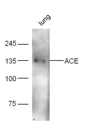
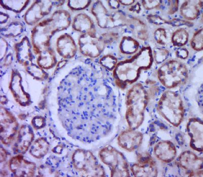
Lung (Mouse) Lysate at 40 ug Primary: Anti-ACE (bs-0439R) at 1/300 dilution Secondary: IRDye800CW Goat Anti-Rabbit IgG at 1/20000 dilution Predicted band size: 147 kD Observed band size: 135 kD  Paraformaldehyde-fixed, paraffin embedded (human kidney tissue); Antigen retrieval by boiling in sodium citrate buffer (pH6.0) for 15min; Block endogenous peroxidase by 3% hydrogen peroxide for 20 minutes; Blocking buffer (normal goat serum) at 37°C for 30min; Antibody incubation with (ACE) Polyclonal Antibody, Unconjugated (bs-0439R) at 1:400 overnight at 4°C, followed by a conjugated secondary (sp-0023) for 20 minutes and DAB staining.
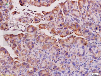
 Tissue/cell: rat pancreas tissue; 4% Paraformaldehyde-fixed and paraffin-embedded;
Antigen retrieval: citrate buffer ( 0.01M, pH 6.0 ), Boiling bathing for 15min; Block endogenous peroxidase by 3% Hydrogen peroxide for 30min; Blocking buffer (normal goat serum,C-0005) at 37∩ for 20 min; Incubation: Anti-ACE1 Polyclonal Antibody, Unconjugated(bs-0439R) 1:200, overnight at 4∑C, followed by conjugation to the secondary antibody(SP-0023) and DAB(C-0010) staining  Cell: Mouse Kidney (4% Paraformaldehyde fixed for 10 minutes ).
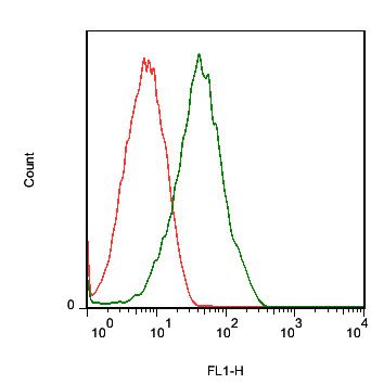
Concentration:1:30. Incubation: 40 minutes. Host/Blank:Mouse Kidney Cells. Flow cytometric analysis of Rabbit Anti-ACE antibody (bs-0439R)(green) compared with control in the absence of primary antibody (red) followed byby Goat Anti-rabbit IgG/FITC antibody (bs-0295G-FITC) secondary antibody . |
